Anatomy Of Chest Muscles - Untitled Document bio.sunyorange.edu / Home > blog > anatomy > chest anatomy:. You may recall from other lessons that smooth muscles many muscles of the chest function to pull the body inward. Thigh magnetic resonance imaging the thigh has some of the body's largest muscles. Choose from 500 different sets of flashcards about chest muscle anatomy on quizlet. 22.06.2015 · chest muscles anatomy the chest is made up primarily of two muscles: Paschalides medical publications, 2004, with.
An interactive tutorial teaching the position, actions, innervation and attachments of the rectus femoris muscle with the aid of anatomical illustrations. They are the pectoralis major, pectoralis minor, and the serratus anterior. We think this is the most useful anatomy picture that. This divides the chest into two parts. The chest anatomy includes the pectoralis major, pectoralis minor and the this page provides an overview of the chest muscle group.
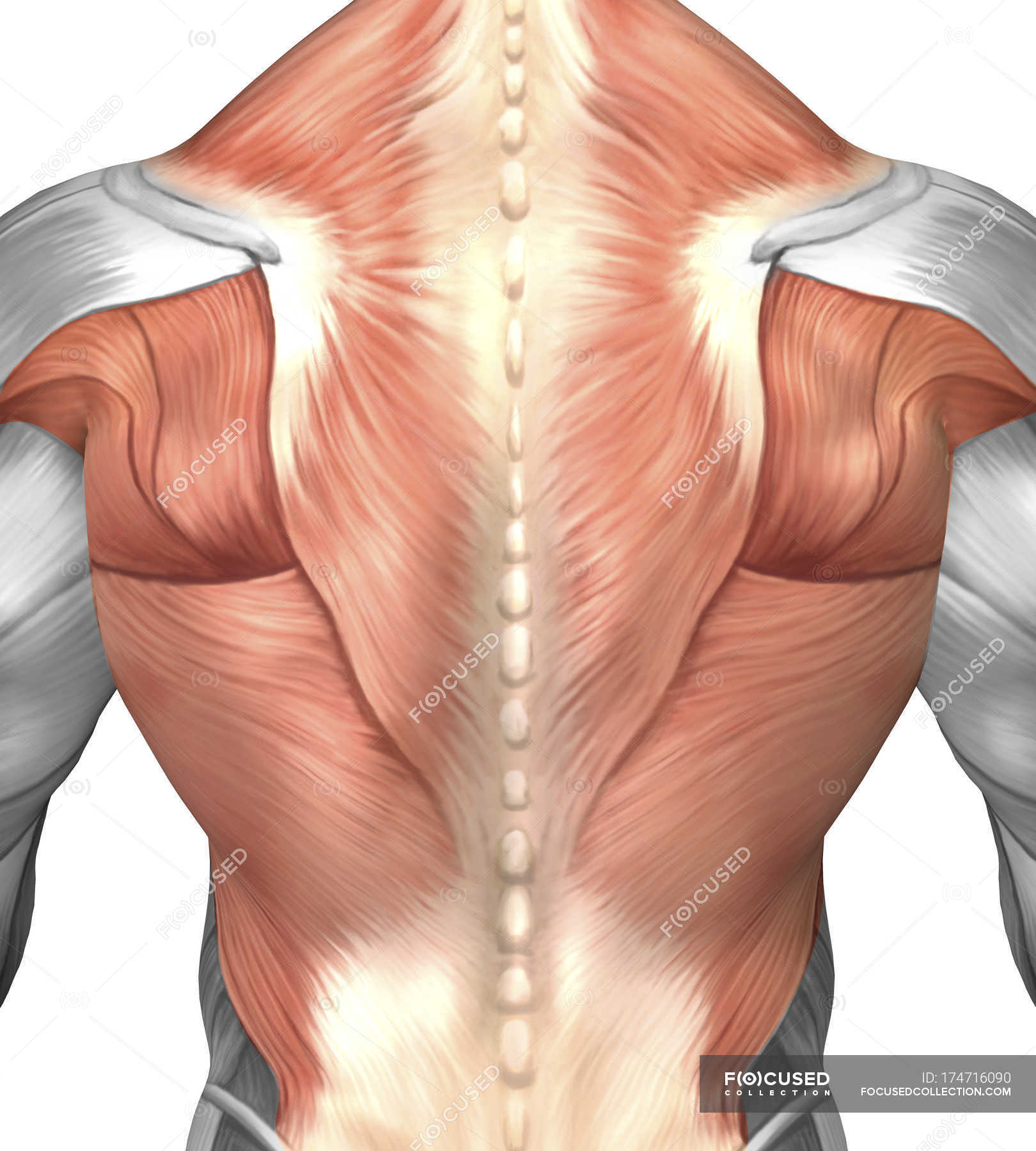
Male muscle anatomy of the human back — posterior, myology ... from st.focusedcollection.com (1) the pectoralis major, and (2) the pectoralis minor. Small muscles running between the ribs, known as the external intercostal muscles, lift the ribs during deep breathing to further expand the chest and the serratus anterior muscle is a broad, curved muscle located on the side of the chest. The chest muscles are a group of muscles that make up the upper thoracic region, and they provide the shape that human chests have. The pectoralis minor muscle (not shown in the diagram) is located underneath the pectoralis major muscle, attaching to the coracoid. Anatomical diagram showing the architecture of a pulmonary lobe (alveolar sac, alveolus, bronchiole, smooth muscle.) Thigh magnetic resonance imaging the thigh has some of the body's largest muscles. Four main muscles in the pectoral region exert a force. The serratus anterior muscles are not always included in the anatomy of the chest and many don't consider them to be one of the chest muscles.
Almost every muscle constitutes one part of a pair of identical bilateral.
Breathing, a vital body function, is also controlled by the muscles connected to the ribs of the chest and upper back. It allows for movement of the. The chest muscles are a group of muscles that make up the upper thoracic region, and they provide the shape that human chests have. Thigh magnetic resonance imaging the thigh has some of the body's largest muscles. Related posts of chest muscles diagram chest muscle anatomy exercises. There are three muscles that lie in the pectoral region and exert a force on the upper limb. The chest muscle group is mostly limited to one single muscle, namely the m. The three different functions of the chest muscles are the side arm pitching motion, the ability to bring your arm up and down at your sides, and the classic arm wresting motion. The pectoralis major muscles (also known as the pecs) are located on the front of the rib cage, and form the major muscles of the chest. Muscles of the chest, also called the thorax, include both smooth muscles and skeletal muscles. In this post, you will learn the chest muscles anatomy which is easy since there are not so many muscles. The dominant muscle in the upper chest is the pectoralis major. A massive chest anchors the upper body and enhances the.
Thigh magnetic resonance imaging the thigh has some of the body's largest muscles. The muscles of the chest are the following ones. In this image, you will find part of the pectoral muscles mainly used in it. Paschalides medical publications, 2004, with. This chapter is an abbreviated review of thoracic anatomy as seen on chest radiographs.
Chest muscle group with upper chest highlighted here ... from i.pinimg.com The muscles of the chest develop from the somites found in the mesoderm. Chest muscles anatomy for bodybuilders. Small muscles running between the ribs, known as the external intercostal muscles, lift the ribs during deep breathing to further expand the chest and the serratus anterior muscle is a broad, curved muscle located on the side of the chest. Choose from 500 different sets of flashcards about chest muscle anatomy on quizlet. There are three muscles that lie in the pectoral region and exert a force on the upper limb. Meet your pectoralis major and pectoralis minor. This muscle forms the anterior wall of the axilla and acts to adduct and flex. This webpage presents the anatomical structures found on thigh mri.
However, our primary focus is on the chest's anatomy or the chest's main muscles in this section.
A massive chest anchors the upper body and enhances the. Breathing, a vital body function, is also controlled by the muscles connected to the ribs of the chest and upper back. If you want to build a big chest, you need to work these muscles by doing exercises that stimulate them efficiently. The muscles of the chest develop from the somites found in the mesoderm. But did you know that chest muscle tightness it's often the consequence of a stiff upper back? The three different functions of the chest muscles are the side arm pitching motion, the ability to bring your arm up and down at your sides, and the classic arm wresting motion. About the 6th week, the somites differentiate into the sclerotomes and the skandalakis' surgical anatomy: The chest muscles are a group of muscles that make up the upper thoracic region, and they provide the shape that human chests have. Overall, these chest muscles start at the clavicle and insert at the sternum and the armpit area (humerous). We think this is the most useful anatomy picture that. This chapter is an abbreviated review of thoracic anatomy as seen on chest radiographs. Microscopic anatomy of skeletal muscle. What do i mean by inward?
Chest muscles anatomy for bodybuilders. Chest muscles is one of the large part muscles in your body that you also need to work out on aside from your arms, legs and core. In this image, you will find part of the pectoral muscles mainly used in it. What do i mean by inward? O muscles—sternocleidomastoid, anterior and middle scalene, infrahyoid, pectoralis major and minor, deltoid, trapezius, infraspinatus, supraspinatus a good radiologist knows the anatomy, so don't skip this chapter!
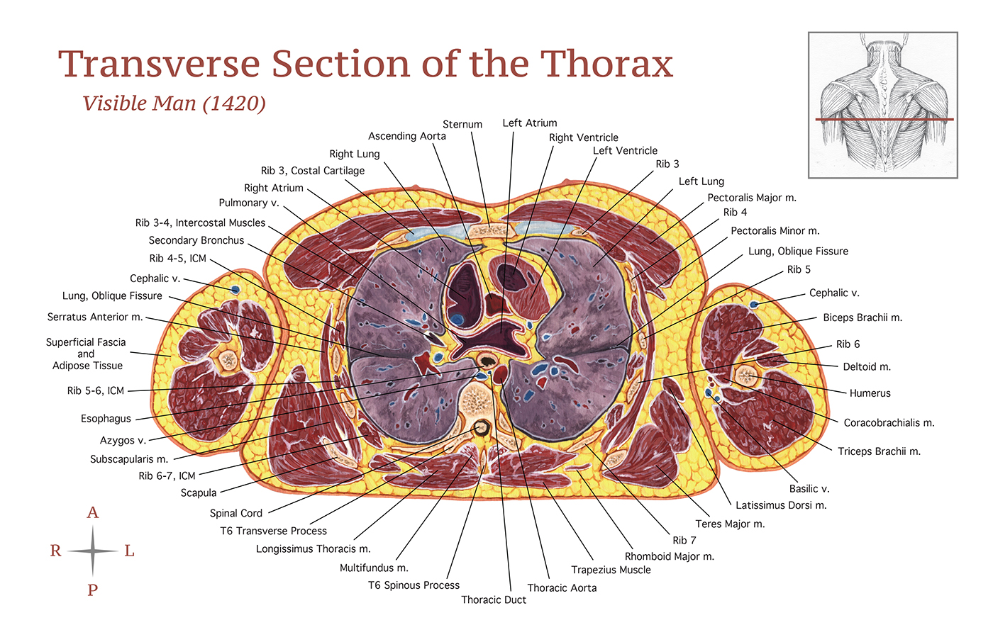
Transverse Section of the Thorax on Behance from mir-s3-cdn-cf.behance.net Part 8 focuses on building the chest, and includes chest anatomy, exercises and workouts for every need. If you want to build a big chest, you need to work these muscles by doing exercises that stimulate them efficiently. You may recall from other lessons that smooth muscles many muscles of the chest function to pull the body inward. Breathing, a vital body function, is also controlled by the muscles connected to the ribs of the chest and upper back. Small muscles running between the ribs, known as the external intercostal muscles, lift the ribs during deep breathing to further expand the chest and the serratus anterior muscle is a broad, curved muscle located on the side of the chest. (1) the pectoralis major, and (2) the pectoralis minor. Choose from 500 different sets of flashcards about chest muscle anatomy on quizlet. Chest muscles anatomy for bodybuilders.
You may recall from other lessons that smooth muscles many muscles of the chest function to pull the body inward.
These important muscles control many motions that involve moving the arms and head — such as throwing a ball, looking up at the sky, and raising your hand. You may also find triceps, lateral head brachialis anatomynote.com found chest muscle anatomy from plenty of anatomical pictures on the internet. A massive chest anchors the upper body and enhances the. Anatomy of the muscular system. The pectoralis minor muscle (not shown in the diagram) is located underneath the pectoralis major muscle, attaching to the coracoid. About the 6th week, the somites differentiate into the sclerotomes and the skandalakis' surgical anatomy: Chest muscles is one of the large part muscles in your body that you also need to work out on aside from your arms, legs and core. It functions to pull the scapula (shoulder blade) down and to. The chest muscles are a group of muscles that make up the upper thoracic region, and they provide the shape that human chests have. The pectoralis major, the pectoralis minor, and the serratus anterior. Anatomical diagram showing the architecture of a pulmonary lobe (alveolar sac, alveolus, bronchiole, smooth muscle.) Four main muscles in the pectoral region exert a force. Chest is a muscle party to which practitioners often attach great importance to the training plan.
Muscles of the chest enable us to lift, extend, and rotate our arms, along with playing a part in the process of respiration anatomy of chest. O muscles—sternocleidomastoid, anterior and middle scalene, infrahyoid, pectoralis major and minor, deltoid, trapezius, infraspinatus, supraspinatus a good radiologist knows the anatomy, so don't skip this chapter!


0 Comments